[ad_1]
Cardiac Magnetic Resonance Imaging (CMRI) segmentation plays a crucial role in diagnosing cardiovascular diseases, particularly ischemic heart conditions, which are a leading cause of global mortality. While CMRI offers precise imaging of anatomical regions with minimal risk, segmentation methods primarily focus on short-axis (SAX) views, leaving long-axis (LAX) views comparatively understudied. However, LAX views are essential for visualizing atrial structures and diagnosing diseases affecting the heart’s apical region, necessitating further exploration and development of segmentation techniques tailored to these views.
State-of-the-art approaches for CMRI segmentation have predominantly concentrated on SAX segmentation using deep learning methods like UNet. Nonetheless, recent advancements, such as the Ω-net method, have started to address the lack of attention on LAX views, utilizing predelineation UNets and Spatial Transformer Networks for orientation normalization and subsequent segmentation. Integrating statistical deformation models and data augmentation techniques like GANs offers promising avenues for improving segmentation accuracy in CMRI, particularly in leveraging the unique advantages of LAX views for comprehensive cardiac imaging and diagnosis. Further research in this domain is essential for enhancing the efficacy of CMRI segmentation in clinical practice.
A new paper by a French research team proposes a robust hierarchy-based augmentation strategy coupled with the Efficient-Net (ENet) architecture for automated segmentation of two-chamber and four-chamber Cine-MRI images. This approach addresses the limitations of previous studies, which have predominantly focused on short-axis orientation, neglecting the intricate structures present in long-axis representations. By leveraging ENet’s efficiency and effectiveness in producing segmentation results with lower computational costs, the research team endeavors to improve segmentation accuracy in long-axis views, particularly in whole-heart segmentation, while also exploring the impact of hierarchical data augmentation on segmentation quality.
The ENet architecture, chosen for its practicality and efficiency, has shown promising results in various medical imaging applications. In this study, the researchers describe the ENet architecture’s adaptation for cardiac Cine-MRI segmentation, specifically focusing on long-axis two- and four-chamber views. Unlike previous works concentrating solely on short-axis segmentation, this research investigates whole-heart segmentation in long-axis views. It evaluates the efficacy of hierarchical data augmentation in improving segmentation accuracy.
The research focuses on producing anatomically accurate segmentation maps through a hierarchy-based augmentation strategy. Two datasets containing Cine-MRI LAX 2-chamber and 4-chamber images were used for training, with specific annotation rules established for each orientation. The ENet architecture, known for its efficiency and effectiveness in segmentation tasks, was adapted for this purpose. The training was conducted on NVIDIA RTX 4500 GPU using the Adam optimizer and a combination loss of multiclass cross-entropy and multiclass Dice. Following a hierarchical procedure involving rotations, intensity alterations, and flipping, data augmentation was employed to improve segmentation accuracy. Evaluation metrics included the Dice coefficient, Hausdorff distance, and clinical metrics such as left ventricular volume and ejection fraction extrapolated from the segmentations. The research highlights the potential of ENet architecture in cardiac MRI segmentation and the importance of hierarchical data augmentation in enhancing segmentation quality.
The results demonstrate notable improvements in segmentation quality, with average Dice and Hausdorff distance enhancements observed. There are also acceptable biases in clinical metric estimation, such as Left Ventricular Ejection Fraction (LVEF). This approach contributes to advancing automated cardiac MRI segmentation and underscores the importance of considering long-axis representations for comprehensive cardiac evaluation.
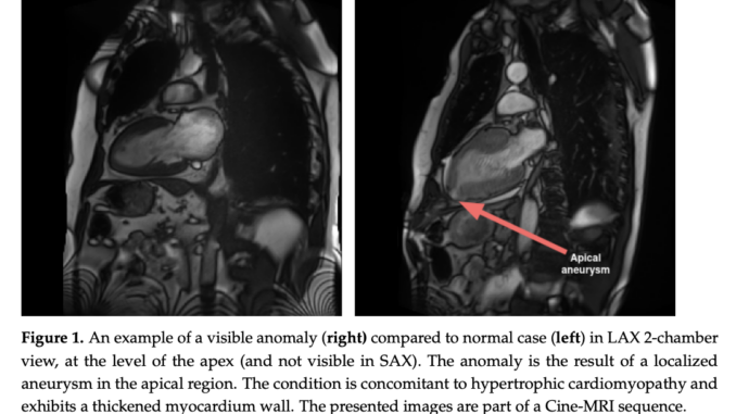
In this research, the research team presents an automated segmentation framework for detecting anatomical structures in Cine-MRI LAX images, which are more complex than SAX orientation. The team’s comprehensive hierarchical data-augmentation strategy produces robust results, even in anomalies and image degradation, enabling accurate computation of the LVEF clinical metric. The ENet CNN architecture shows promise for whole-heart segmentation in two- and four-chamber sequences, offering compact sizes suitable for real-time applications. Although some precision loss near anatomical frontiers was noted, the segmentation quality supports its clinical utility. Additionally, a comparison with a barebone UNet architecture revealed comparable performance, suggesting potential for further optimization.
Check out the Paper. All credit for this research goes to the researchers of this project. Also, don’t forget to follow us on Twitter and Google News. Join our 37k+ ML SubReddit, 41k+ Facebook Community, Discord Channel, and LinkedIn Group.
If you like our work, you will love our newsletter..
Don’t Forget to join our Telegram Channel
![]()
Mahmoud is a PhD researcher in machine learning. He also holds abachelor’s degree in physical science and a master’s degree intelecommunications and networking systems. His current areas ofresearch concern computer vision, stock market prediction and deeplearning. He produced several scientific articles about person re-identification and the study of the robustness and stability of deepnetworks.
[ad_2]
Source link


Be the first to comment